Label-free visualization of ciliary rootlets in mouse brain


Label-free visualization of ciliary rootlets in mouse brain
Murakami, Y.; Nuriya, M.; Hu, Z.; Tomioka, M.; Oketani, R.; Hiramatsu, K.; Leproux, P.; Inoko, A.; Honjoh, S.; Kano, H.
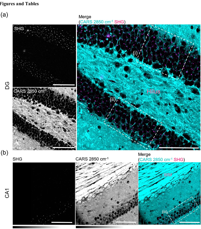
AbstractNeuronal primary cilia are important role in brain development, sensory perception and neurogenesis. Rootletin, a fibrous protein composed of coiled-coil motifs, is a major structural component of ciliary rootlets and is essential for understanding ciliary functions. However, the precise mechanisms by which Rootletin influences ciliary dynamics and impacts neuronal function remain largely unknown, primarily due to the challenges in visualizing these structures. Here, we describe a label-free, rapid, and highly sensitive method to visualize Rootletin molecules in brain tissue. This platform integrates a second harmonic generation (SHG) microscope and background reduction by a one-step chemical pretreatment. Additionally, we employ coherent anti-Stokes Raman scattering imaging to simultaneously determine the cellular regions and intracellular locations of SHG signals. By applying this multimodal multiphoton imaging to mouse hippocampus, we found that neuronal ciliary rootlets were found to exhibit highly organized specific intracellular distributions. Moreover, the formation of ciliary rootlets precedes that of primary cilia. These findings highlight the utility of our label-free imaging platform in developmental and neuroscience research, providing a new tool to characterize ciliary dynamics and neuronal function.