Partitioning of nuclear material into apoptotic fragments through establishment of asymmetric cell death morphology

Partitioning of nuclear material into apoptotic fragments through establishment of asymmetric cell death morphology
Santavanond, J. P.; Jiang, L.; Hodge, A. L.; Ozkocak, D. C.; Ceviker, A.; Arakawa, S.; Shimizu, S.; Yoshino, I.; Rutter, S. F.; Phan, T. K.; Tixeira, R.; Baxter, A. A.; Caruso, S.; Newton, L. M.; Stephens, R.; Humbert, P. O.; Hulett, M. D.; Atkin-Smith, G. K.; Poon, I. K.
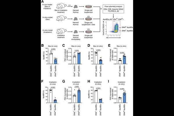
AbstractCellular material in apoptotic cells must be efficiently cleared by phagocytes to maintain tissue homeostasis. Defects in this process can lead to the onset of secondary necrosis and the release of intracellular contents such as damage associated molecular patterns (DAMPs) and autoantigens that are often derived from the nucleus. Therefore, appropriate handling and clearance of apoptotic material is vital to prevent unwanted inflammatory response and the onset of autoimmune disorders. However, how nuclear material is packaged by apoptotic cells for effective clearance by phagocytes is not well understood. By utilising murine models of apoptosis, we observed that a distinct subset of large extracellular vesicles generated from apoptotic thymocytes, known as apoptotic bodies (ApoBDs), can harbour the majority of nuclear contents. Mechanistically, we discovered that apoptotic cells can asymmetrically partition the nucleus into a single large membrane bleb located at one side of the cell, with other cellular contents such as mitochondria and acid organelles distributed to the opposite side. Whilst this newly observed apoptotic morphology, coined as asymmetric cell death morphology (AsyCDM), is morphologically similar to the process of erythroblast enucleation, pharmacological compounds that could interfere with erythroblast enucleation did not block the establishment of AsyCDM during apoptosis. Notably, AsyCDM was reliant on the contractile forces generated by ROCK1-dependent plasma membrane blebbing. Taken together, this study suggests that intracellular contents are partitioned into different ApoBD subsets during apoptosis through a regulated process driven by ROCK1-dependent actomyosin contraction.