Multiscale Biomechanical and Electrophysiological Modeling of Nociceptive Activation in Musculoskeletal Joint Disorders: Insights from the Temporomandibular Joint

Multiscale Biomechanical and Electrophysiological Modeling of Nociceptive Activation in Musculoskeletal Joint Disorders: Insights from the Temporomandibular Joint
Chen, J.; Sun, S.; Ahmadi, F.; Chen, P.; Chai, J.; Zhao, J.; Damon, B.; Almpani, K.; Lee, J.; Yao, H.
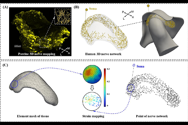
AbstractBackground and objective: Musculoskeletal joint disorders often show inconsistent relationships between structural degeneration and nociceptive pain. Temporomandibular joint (TMJ) disc displacement represents a clinically relevant model for investigating the structure-function-pain relationship. This study aimed to develop a multiscale computational framework integrating biomechanics, three-dimensional (3D) neural morphology, and electrophysiology to quantitatively link TMJ structural alterations, biomechanical loading, and peripheral nociceptive activation. Methods: Strain distributions in the TMJ disc and retrodiscal tissue during mouth opening and clenching were computed in ArtiSynth under varying degrees of displacement. Human TMJ 3D nerve architecture was reconstructed using porcine TMJ nerve mapping data as an anatomical reference, and ion channel dynamics were implemented in NEURON. Model coupling was achieved by mapping biomechanical strain fields onto nociceptor membranes to simulate mechanosensitive currents and action potential propagation to the trigeminal ganglion. Results: Anterior DDwoR induced a severity dependent strain pattern in the TMJ disc and retrodiscal tissue, including posterior redistribution, increased strain magnitude, prolonged activation, and broader retrodiscal tissue involvement. Displacements of 4, 6, and 8 mm produced larger mechanosensitive currents, broader terminal depolarization, and higher trigeminal firing rates during mouth opening (6, 18, and 28 Hz) and clenching (8, 20, and 28 Hz), whereas 0- and 2-mm displacements produced negligible neural activation. Conclusions: This study establishes a multiscale biomechanical-electrophysiological framework linking TMJ structural alterations to peripheral nociceptive activation. The framework quantitatively connects macroscale strain patterns with microscale neural activation, suggesting that anterior disc displacement may amplify peripheral nociceptive signaling by increasing the overlap between elevated strain and densely innervated retrodiscal tissue.